MAT 594 - 2008S
Melissa Carrasco - Imaging and Biological Research
At UCSB, the Retinal Biology Lab and the Center for Bio-Image Informatics work together to further research in the area of retinal detachment and reattachment. The research goal of the Retinal Cell Biology Lab is to understand the cellular changes that occur following retinal detachment and reattachment. One way this is done is by observing images of retinal cells that are obtained via confocal microscopy.
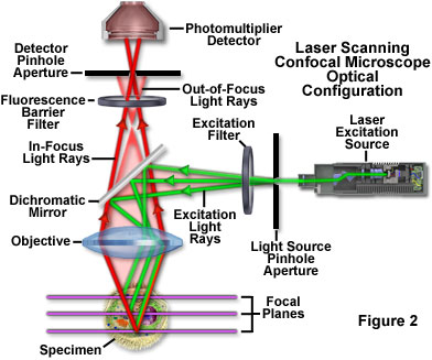
Confocal microscopy is an optical imaging technique that uses point illumination. One point of the specimen is illuminated at a time. The specimen is treated with fluorescent dye and as it is illuminated, it will emit a certain color depending on the color of light that is used to illuminate and the properties of the cells of the specimen at that point. The emitted light is captured and the information is used to form a pixel of the image. [4]
 [3]
[3]
To get a complete visualization of the specimen, multiple passes using a different color light will be used to scan the specimen. Each pass will create will create a separate image and the images will be later merged together using software. Below is a confocal image of a cat retina that has been detached for seven days. Additional images can be found on the Retinal Cell Biology website. [4]
Image segmentation is used to automatically find the boundaries of the retinal layers in the image. This allows for the viewer to easily get an overview of the shape of the layer. [2]
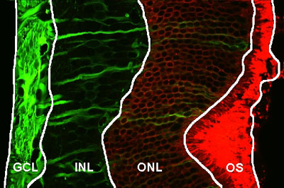
GCL: ganglion cell layer, INL: inner nucleus layer, ONL: outer nucleus layer, OS: rod outer segment
Once the cell layer has been identified, cell nuclei can be automatically identified and marked, which makes it easier for the viewer to identify individual cells.
[2]Center for Bio-Informatics
[3]Introduction to Confocal Microscopy
[4]Confocal Microscopy