MAT 594 - 2008S
Pablo Colapinto - Qdot Imaging
This preliminary research survey is based on work being conducted by affiliates of the CNSI, namely Shimon Weiss, Faculty Director, and Laurent A. Bentolila, Scientific Director of the Advanced Microscopy and Spectroscopy lab UCLA. According to Weiss:
"The unique optical properties of qdots enable to multiplex many different biological signals in complex environments such as the living cell."
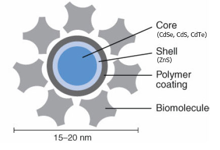
Qdots, or Quantum Dots, are semiconductor groups, or 'puddles', of electrons. Each is about 20-200 nanometers in diameter (billionths of a meter), though they vary in size.
Sources of all imagery is from a pdf circulated by Invitrogen, unless otherwise noted
Their manipulation is on the forefront of nanotechnology research, with applications ranging from bioimaging to computation. This research focusses on its use in bioimaging, which is currently an active application of Qdots. It should be noted, however, that their 'qubit' computational potential is not a separate consideration. Rather, it is precisely their ability to be functional - i.e. to be turned on -- that suggests they will revolutionize microscopy.
Here we explore Qdots as a non-organic fluorescent semiconductor. Compared to organic protein-based fluorescent imaging, Qdot Imaging offers the following major benefits:
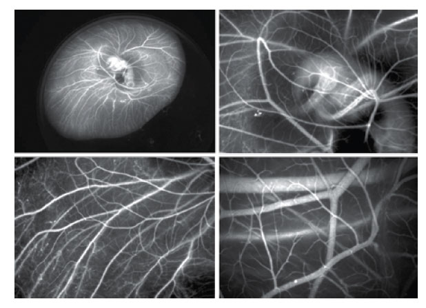
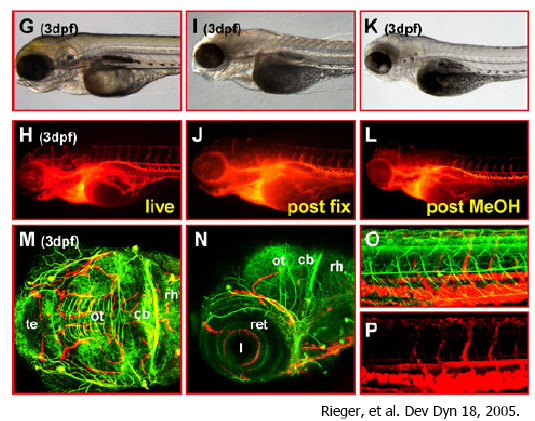
1 - Potential to work at Macro, Micro, and Nanoscales, all within one probe.
Here is an Qdot image of the vascular structure of a baby chick at multiple resolutions.
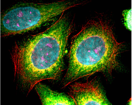
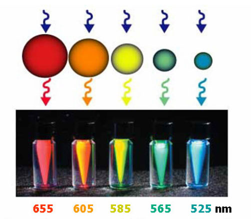
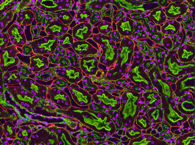
2 - Broad excitation spectrum and narrow emission spectrum. Depending on their size, their confinement can manipulated in one or more dimensions, making them emit very specific colors. This enables "multiplex" coding, something difficult with traditional protein-based dyes. Here is the first five-color cell image ever produced, made with Qdot imaging techniques.
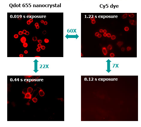
3 - Very sensitive - High Signal-to-Noise Ratio. They are "brighter" because of a high quantum yield (every photon absorbed produces a large emission in response).
4-Can be imaged in vivo -- in live specimens.
5 - Can be tracked, in real-time, in living tissue, over a long period of time (~ 15 hours . . .)
Qdots can be "confined" in their excitation by varying their size. This means they can be forced to emit specific colors, even while remaining sensitive to a wide range of light.
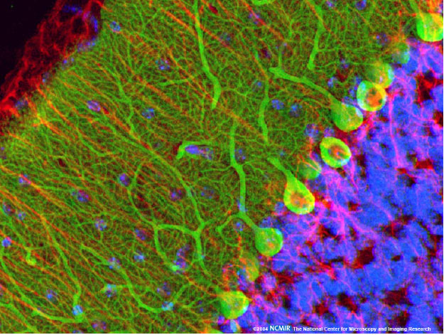
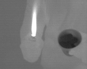
Nikon has launched competitions seeking the best nanoimages -- Thomas Deerinck at the NIMR of UCSD (see Resouces section below) is reknown for his microscopy images, including this one of a mouse liver:
Qdots could be easily be made sensitive to electric and magnetic Fields
"Functionalizations" include the possibility of built-in on/off switches, Photoelectric biotransducers, and other robotic like controls to activate the image.
A final note on computing with Qubits: Quantum entanglement, the process by which the "spin" of two quantum dots are intricately connected, is a promising source of 'qubit' computations. Indeed, each 'qubit' should be able to hold two values simultaneously. Einstein famously denounced entanglement, calling it "Spooky Action at a Distance."
Invitrogen Qdot Imaging Company
Nikon Small World Photo Competition
Qdot Imaging Labs in the UC System:
CNSI @ UCLA Core Facilities Description
Electron Imaging Center for NanoMachines
at the University of California, San Diego
(2) S. Weiss, et al. "Advances in fluorescence imaging with quantum dot bio-probes", Biomaterials 27 (2006) 1679Ð1687
(3) H. Arya et al. "Quantum dots in bio-imaging: Revolution by the small", Biochemical and Biophysical Research Communications 329 (2005) 1173Ð1177
(4) "The future of fluorescence Qdot nanocrystal technology", invitrogen company brochure.